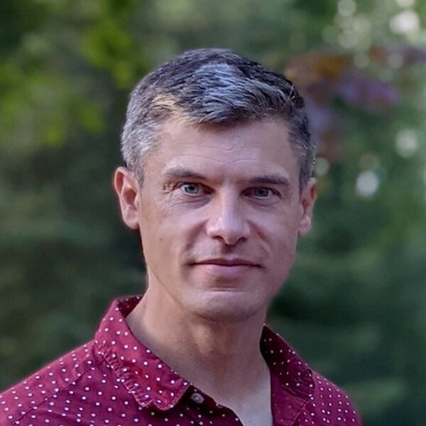
Bradley MacIntosh
PhD, University of Toronto

At A Glance
Our primary desire is to utilize non-invasive brain imaging techniques to study chronic diseases through a vascular lens with the goal of identifying new therapies. Our focus is on computational methods, exploiting artificial intelligence deep machine learning, to automate and characterize brain diseases. In summary, the novel use of existing tools and the development of new techniques aim to improve our current understanding of brain health, resilience, treatment, and recovery.
Short Bio
Bradley MacIntosh is a Senior Scientist within the Hurvitz Brain Sciences program & Physical Sciences platform at Sunnybrook Research Institute. MacIntosh is a member of the Sandra E. Black Centre for Brain Resilience and Recovery and was a member of the Heart and Stroke Foundation Canadian Partnership for Stroke Recovery. MacIntosh is a Professor in the Department of Medical Biophysics at the University of Toronto, which is also where he completed his PhD in 2006 (supervision by Simon Graham). MacIntosh did a MSc at the Robarts Research Institute at Western University (supervision by Ravi Menon), and a postdoctoral fellowship at the University of Oxford’s Centre for Functional Magnetic Resonance Imaging of the Brain (FMRIB; supervision by Peter Jezzard). Over the last decade, the lab has worked collaboratively to create diverse external research teams, including ongoing work with CAMH in Toronto and the Computational Radiology & Artificial Intelligence unit in Oslo, Norway.
Research Synopsis
The MacIntosh lab focuses on developing and translating vascular imaging tools and measures to advance our understanding of human brain disease. Magnetic Resonance Imaging (MRI) is a versatile medical imaging modality, and we use this method for functional imaging to study neurovascular function, cerebral blood flow, and related aspects of brain physiology and pathophysiology.
METHODS: Our primary method of Arterial Spin Labeling (ASL) magnetic resonance technique is predominant in our research activities. This technique produces non-invasive cerebral blood flow maps with spatial resolution that approaches the millimeter details of a conventional anatomical picture. Our ASL innovation includes image processing of individual scans, a pipeline to use these images in group analysis or multi-site trials, and additional ASL hemodynamic features (such as the arterial transit time and the spatial coefficient of variation). On-going efforts arousing deep learning to improve physiological MRI in radiology applications.
CLINICAL MOTIVATION: With our focus on physiological and pathophysiological imaging techniques, the desired outcome is to help patient populations in the areas of stroke, small vessel disease, Alzheimer’s disease, bipolar disorder, and diabetes. For instance, the MacIntosh lab, in collaboration with Oslo University Hospital, has applied deep-segmentation neural networks in various neuroradiological applications (i.e., gliomas, white matter hyperintensities from small vessel disease, intracranial hemorrhages), enabling the creation of predictive models for disease progression and providing valuable metrics to support clinical decision-making. We also examine cerebral blood flow pattern differences and quantifiable features as a potential biomarker to better understand clinical populations such as youth with bipolar disorder and those affected by genetic frontotemporal dementia.
COLLABORATION: The MacIntosh lab has a focus on clinical translation and work with Clinician-Scientist colleagues. The lab is involved in a variety of internal, national, and international initiatives which includes a national drug trial through the Canadian Partnership for Stroke Recovery, an internal clinical trial through the Harquail Centre for Neuromodulation examining the efficacy and brain network connectivity changes of psilocybin-assisted psychotherapy on major depressive disorder, an international LeDucq Foundation network to study perivascular spaces in small vessel disease and the detrimental effects of sleep apnea on the brain. The lab is also focused on developing deep learning tools to characterize brain diseases at the Computational Radiology & Artificial Intelligence unit (CRAI.no) at the Oslo Hospital University.
Recent Publications
- MacIntosh B. J., Liu, Q., Schellhorn, T., Beyer, M. K., Groote, I. R., Morberg, P. C., Poulin, J. M., Selseth, M. N. Bakke, R. C., Naqvi, A., Hillal, A., Ullberg, T., Wassélius, J., Rønning, O. M., Selnes, P., Kristoffersen, E. S., Emblem, K. E., Skogen, K., Sandset, E. C., & Bjørnerud, A. (2023, September 28). Radiological features of brain hemorrhage through automated segmentation from computed tomography in stroke and traumatic brain injury. Frontiers in Neurology, 14, 1244672.
- Luciw, N. W., Grigorian, A., Dimick, M. K., Jiang, G., Chen, J. J., Graham S. J., Goldstein, B. I., & MacIntosh, B. J. (2023, August 29). Classifying youth with bipolar disorder versus healthy young using cerebral blood flow patterns. Journal of Psychiatry and Neuroscience, 48(4), E305-E314.
- Valsamis, J. J., Luciw, N. J., Haq, N., Atwi, S., Duchesne, S., Cameron, W., MacIntosh B. J. (2023, July). An imaging-based method of mapping multi-echo BOLD intracranial pulsatility. Magnetic Resonance in Medicine, 90(1), 343-352.
- Kim, W. S. H., Xiang, J., Roudala, E., Chen, J. J., Gilboa, A., Sekuler, A., Gao, F., Lin, Z., Jegatheesan, A., Masellis, M., Goubran, M., Rabin, J. S., Lam, b., Cheng, I., Fowler, R., Heyn, C., Black, S. E., Graham, S. J., & MacIntosh B. J. (2023, August). MRI assessment of cerebral blood flow in nonhospitalized adults who self-isolated due to COVID-19. Journal of Magnetic Resonance Imaging, 58(2), 593-602.
- Luciw, N. J., Shirzadi, Z., Black, S. E., Goubran, M., & MacIntosh, B. J. (2022, February 19). Automated generation of cerebral blood flow and arterial transit time maps from multiple delay arterial spin-labeled MRI. Magnetic Resonance in Medicine, 88(1), 406-417.
Visit the Google Scholar profile of Dr. Bradley MacIntosh.
Graduate Students
Current graduate students
Guocheng Jiang
Information for prospective graduate students
Desirable aspects found within strong applicants to the MacIntosh Lab include:
- A passion for the lab’s research areas of interest which may include one or more of applied advanced neuroimaging techniques, AI-driven computational neuroradiology, and clinical populations that include stroke, small vessel disease, diabetes, dementia, Alzheimer’s, and bipolar disorder.
- An academic record that includes computational methods and/or biostatistics
- Previous research experience in a medical biophysics field or related field
- Independent research experience that may include a thesis or dissertation, poster presentations, publication experience, etc.
