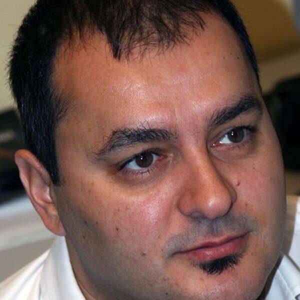
Kâmil Uludağ
PhD, Humboldt Universität

At a Glance
- MRI sequences at 3, 7 and 9.4 Tesla human scanners
- Physical and physiological foundations of functional MRI
- Clinical application of quantitative MRI at 3 and 7 Tesla
- Anatomical MRI in post mortem and in vivo brain tissue
- Brain blood perfusion using arterial spin labelling MRI
- Neuroscience in healthy subjects and patients
- Artificial intelligence in fundamental Neuroscience and Diseases
Appointments
Techna Institute & Koerner Scientist in MR Imaging, Joint Department of Medical Imaging and Krembil Brain Institute
Short Bio
Kâmil Uludağ studied from 1992 till 1997 Physics at the Technical University of Berlin. He completed his Ph.D. in Physics in 2003 on Near-Infrared Optical Spectroscopy (Humboldt University, Berlin) and moved for a postdoc position to the Center for Functional MRI (UCSD, San Diego, USA) to work on the physiological and physical basis of functional MRI. In 2004, he was appointed Head of Human Brain Imaging group at the Max-Planck-Institute for Biological Cybernetics, Tübingen. From June 2010 to December 2018, he was Associate Professor in the Faculty of Psychology & Neuroscience and Head of the Department of Cognitive Neuroscience continuing his work on the basis of fMRI utilizing the new Ultra-High Field human MRI scanners (7 and 9.4 Tesla). Since May 2019, he is Full Professor at the Department of Medical Biophysics, University of Toronto.
Dr. Uludağ is on the editorial board of five neuroimaging journals, served from 2011 to 2013 as Annual Meeting Committee Member of the International Society for Magnetic Resonance Imaging in Medicine (ISMRM) and was elected Chair of the Current Issues of Brain Function study group. He recently edited a textbook “functional MRI: from Nuclear Spins to Brain Functions” (Publisher: Springer).
Research Synopsis
Magnetic resonance imaging (MRI) at 1.5, 3 and 7 Tesla magnetic field strength is an excellent tool to image soft tissue in the brain and body for fundamental research and clinical applications. However, the relationship between MRI contrasts and the underlying biochemistry, connectivity and cognitive processes is often not clear and currently limits the interpretation of MRI data.
Dr. Uludağ’s laboratory combines artificial intelligence approaches with 1.5 and 3T MRI big data in order to answer fundamental neuroscience questions and to develop biomarkers for clinical applications. Furthermore, he uses deep learning methods and generative models to improve the effectivity of MR image acquisition and reconstruction, promising to advance our understanding of the physical and physiological basis of MRI. As Co-Director of the Slaight Family Centre for Advanced MRI at the Toronto Western Hospital, Dr. Uludağ advises the clinical research groups in their studies of the brain and spine.
In addition, the research interests of Dr. Uludağ’s laboratory include studying cognition and anatomy in the human brain using Ultra-High Field human MRI scanners (7 and 9.4 Tesla). Ultra-high field MRI is an enabling technology that is increasingly used by researchers and clinicians for human neuroimaging to ask novel questions about brain structure and function. His lab works on quantitative anatomical and functional MRI methods (for example, ASL, T1, T2*, and SWI) and applies these cutting-edge approaches on post mortem brains, healthy subjects and patients. Dr. Uludağ’s group will continue to work on 7T MRI data acquired at national and international MRI centres (specifically in Montreal, London (Ontario), Suwon (South Korea), Boston, Minneapolis and Maastricht) and perform advanced data analysis (including AI and deep learning) on this data, which is unprecedented in spatial resolution, contrast and information content.
Recent Publications
- J. Poublanc, O. Sobczyk, R. Shafi, J. Duffin, K. Uludağ, J. Wood, C. Vu, R. Dharmakumar, J.A. Fisher, D.J. Mikulis. Perfusion MRI using endogenous deoxyhemoglobin as a contrast agent: preliminary data. Magnetic Resonance in Medicine (accepted, 2021).
- S.H. Han, S. Eun, H. Cho, K. Uludağ, S.G. Kim. Improvement of sensitivity and specificity for laminar BOLD fMRI with double spin-echo EPI in humans at 7 T. NeuroImage (accepted, 2021)
- S. Kashyap, D. Ivanov, M. Havlicek, L. Huber, B.A. Poser, K. Uludağ. Sub-millimetre resolution laminar fMRI using Arterial Spin Labelling in humans at 7 T. Plos One (accepted, 2021).
- H.M. Wiesner, D.Z. Balla, K. Scheffler, K. Uğurbil, X. Zhu, W. Chen, K. Uludağ*, R. Pohmann*. Quantitative and simultaneous measurement of oxygen consumption rates in rat brain and skeletal muscle using 17O MRS imaging at 16.4T. Magnetic Resonance in Medicine (accepted, 2020). * equal contribution
- N.Priovoulos, S.C.J. van Boxel, H.I.L. Jacobs, B.A. Poser, K. Uludag, F.R.J. Verhey, D. Ivanov. Unravelling the contributions to the neuromelanin-MRI contrast. Brain Structure and Function (accepted, 2020).
- R.A.M. Haast, J. Lau, R. Menon, D. Ivanov, K. Uludağ, A. Khan. Effects of MP2RAGE B1+ sensitivity on inter-site T1 reproducibility and hippocampal morphometry at 7T. NeuroImage (2020, accepted).
- J. Lau, Y. Xiao, R.A.M. Haast, G. Gilmore, K. Uludag, K. MacDougall, R. Menon, A. Parrent, T. Peters, A. Khan. Direct visualization and characterization of the human zona incerta and surrounding structures. Human Brain Mapping 41: 4500-4517 (2020).
- L. Huber, E.S. Finn, Y. Chai, R. Goebel, R. Stirnberg, T. Stöcker, S. Marrett, K. Uludag, S.G. Kim, S. Han, P.A. Bandettini, B.A. Poser. Layer-dependent functional connectivity methods. Prog Neurobiol. (accepted, 2020).
- H.I.L. Jacobs, N. Priovoulos, B.A. Poser, L.H.G. Pagen, D. Ivanov, F.R.J. Verhey, K. Uludag. Dynamic behavior of the locus coeruleus during arousal-related memory processing: a multi-modal 7T fMRI study. eLife (2020).
- I. Marquardt, M. Schneider, O.F. Gulban, D. Ivanov, P. De Weerd, K. Uludağ. Feedback Contribution to Surface Motion Perception in the Human Early Visual Cortex. eLife (2020).
- J.P.D. Kleinloog, R.P. Mensink, D. Ivanov, J.J. Adam, K. Uludag, J.P. Joris. Aerobic Exercise Training Improves Cerebral Blood Flow and Executive Function: A Randomized, Controlled Cross-Over Trial in Sedentary Older Men. Frontiers in Aging Neuroscience (2019).
- M. Havlicek, K. Uludağ. A Dynamical Model of the Laminar BOLD Response. NeuroImage 204 (2019).
- E. Düzel, J. Acosta-Cabronero, D. Berron, G.J. Biessels, I. Björkman-Burtscher, M. Bottlaender, R. Bowtell, M. v Buchem, A. Cardenas-Blanco, D. Chan, S. Clare, A. Fillmer, P. Gowland, O. Hansson, J. Hendrikse, O. Kraff, I. Ronen, E. Petersen, J. Rowe, H. Siebner, T. Stoeker, M. Tosetti, K. Uludag, A. Vignaud, J. Zwanenburg, O. Speck. European Ultrahigh-Field Imaging Network for Neurodegenerative Diseases (EUFIND). Alzheimer's & Dementia: Diagnosis, Assessment & Disease Monitoring 11, 538-549 (2019).
- M.J. Betts, E. Kirilina, M. Otaduy, D. Ivanov, J. Acosta-Cabronero, M. Callaghan, C. Lambert, A. Cardenas-Blanco, K. Pine, L. Passamonti, C. Loane, M.C. Keuken, P. Trujillo, F. Lüsebrink, H. Mattern, K. Liu, N. Priovoulos, K. Fließbach, M.J. Dahl, A. Maaß, C.F. Madelung, D. Meder, A.J. Ehrenberg, O. Speck, N. Weiskopf, R. Dolan, B. Inglis, D. Tosun, M. Morawski, F.A. Zucca, H.R. Siebner, M. Mather, K. Uludag, H. Heinsen, B.A. Poser, R. Howard, L. Zecca, J.B. Rowe, L.T. Grinberg, H.I.L. Jacobs, E. Düzel, D. Hämmerer. Locus coeruleus imaging as a biomarker for noradrenergic dysfunction in neurodegenerative diseases. Brain 142, 2558-71 (2019).
- L. Huber, K. Uludağ*, H. E. Moeller*. Non-BOLD contrast for laminar fMRI in humans: CBF, CBV, and CMRO2. NeuroImage 197, 742-760 (2019).* equal contribution
- J. Polimeni, K. Uludağ. Neuroimaging with Ultra-High Field MRI: Present and Future. NeuroImage 168, 1-532,. (2018, Publisher: Elsevier).
- K. Uludağ, K. Ugurbil, L. Berliner. Functional MRI: From Nuclear Spins to Brain Function. (2015, Publisher: Springer).
- K. Uludağ, P. Blinder. Linking brain vascular physiology to hemodynamic response in ultra-high field MRI. NeuroImage 168, 279-295 (2018).
- S. Kashyap, D. Ivanov, M. Havlicek, S. Sengupta, B.A. Poser, K. Uludağ. Resolving laminar activation in human V1 using ultra-high spatial resolution fMRI at 7T. Scientific Reports 8, 1-11 (2018).
- I. Marquardt, M. Schneider, O.F. Gulban, D. Ivanov, K. Uludağ. Cortical depth profiles of luminance contrast responses in human V1 and V2 using 7 T fMRI. Human Brain Mapping 39, 2812-27 (2018).
- M. Havlicek, A. Roebroeck, K. J. Friston, A. Gardumi, D. Ivanov, K. Uludağ. Physiologically informed dynamic causal models for fMRI. NeuroImage 122, 355-372 (2015).
- R.A.M. Haast, D. Ivanov, R.J.T. IJsselstein, S.C.E.H. Sallevelt, J.F.A. Jansen, H.J.M. Smeets, I.F.M. de Coo, E. Formisano, K. Uludağ. Anatomic & metabolic brain markers of the m.3243A>G mutation: A multi-parametric 7T MRI study. NeuroImage Clinical 18, 231-244 (2018).
- M. Havlicek, D. Ivanov, A. Roebroeck, K. Uludağ. Determining excitatory and inhibitory neuronal activity from fMRI data using generative model. Frontiers in Neuroscience 11, Article 616 (2017).
- K. Uludağ. To dip or not to dip: reconciling optical imaging and fMRI data. Proc Natl Acad Sci U S A 107(6), E23 (2010).
- K. Uludağ, B. Müller-Bierl, K. Ugurbil. An integrative model for neuronal activity-induced signal changes for gradient and spin echo functional imaging. Neuroimage 48(1), 150-65 (2009).
Graduate Students
Fatemeh Bagheri
Shawn Carere
Angelica Manalac
Sayan Nag
Brian Nghiem
Jacob Schulman
Labeeb Talukder
