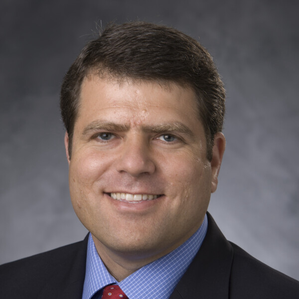
David Kirsch
MD, PhD, Johns Hopkins School of Medicine

At A Glance
- As a clinician scientist sarcoma radiation oncologist, our lab uses genetically engineered mouse models of sarcoma with complimentary human sarcoma cell lines and tissues to study sarcoma biology and radiation biology.
- We study mechanisms of sarcoma development and tumour maintenance in undifferentiated small round cell sarcoma driven by the CIC-DUX4 fusion oncoprotein and in epithelioid sarcoma initiated by loss of SMARCB1 a subunit of the SWI/SNF protein complex.
- We also study mechanisms of tumour suppression by the three genes most frequently mutated in complex karyotype sarcomas like undifferentiated pleomorphic sarcoma: p53, Rb, and ATRX
- Using autochthonous sarcoma models, we study mechanisms of tumour response to radiotherapy alone or radiation and immunotherapy
- We use Cre-loxP technology to delete genes in a cell-type specific manner and apply sophisticated small animal irradiation to study mechanisms of radiation injury and regeneration in the intestine and other tissues.
Short Bio
David Kirsch, MD, PhD, is a Senior Scientist at Princess Margaret Cancer Centre and the Peter and Shelagh Godsoe Chair in Radiation Medicine. After graduating from Duke University with a BS in Biology, he completed the MD/PhD program at Johns Hopkins School of Medicine, where he performed his thesis research with Dr. Michael Kastan. Dr. Kirsch completed residency training in radiation oncology at Massachusetts General Hospital and a post-doc in the laboratory of Dr. Tyler Jacks at MIT. In 2007 Dr. Kirsch returned to Duke, to establish an independent research program. In 2023, Dr. Kirsch was recruited to Princess Margaret Cancer Centre at University Health Network. He has received a number of awards for his research including the 2010 Michael Fry Award and the 2017 J.W. Osborne Award from the Radiation Research Society. Dr. Kirsch has been elected to the American Society for Clinical Investigation, the Association of American Physicians, a Fellow of the American Society for Radiation Oncology, and a Fellow of the American Association for the Advancement of Science. He has also received several awards for mentoring including the 2014 Duke University Dean’s Award for Excellence in Mentoring and the 2021 Career Mentoring Award in Basic/Translational Science from the Duke University School of Medicine.
Research Synopsis
Our laboratory utilizes sophisticated genetically engineered mouse models (GEMMs) to study mechanisms of sarcoma development and the response of tumours and normal tissues to radiation therapy alone or in combination with immunotherapy. Sarcomas are mesenchymal tumours of the connective tissues, which include muscle, fat, blood vessels, and bone. Approximately one-third of sarcomas have a simple karyotype with a primary genetic driver, such as a fusion oncoprotein like EWS-FLI or FUS-CHOP. The remaining sarcomas have complex karyotypes and are characterized by mutations in tumour suppressor genes like p53, Rb, and ATRX, which are also mutated in many other cancers.
Using a combination of genetically engineered mouse models, mouse and human cell lines, and human samples, we use molecular and cell biology to study mechanisms of sarcoma development, metastasis, and response to therapy. Current projects utilize cutting-edge tools in molecular biology, genetics, genomics, and imaging to study sarcomas initiated by the CIC-DUX4 fusion protein, loss of SMARCB1, and loss of the tumour suppressor genes p53, Rb, and ATRX. Studying the role of p53 in cancer development and the response of normal tissues to radiation therapy has been a long-standing interest to our lab. Ongoing projects study non-canonical roles of p53 in regulating transposable elements and 3D DNA topology as potential mechanisms of tumour suppression.
Recent Publications
- Wisdom AJ, Mowery YM, Hong CS, Himes JE, Nabet BY, Qin X, Zhang D, Chen L, Fradin H, Patel R, Bassil AM, Muise ES, King DA, Xu ES, Carpenter DJ, Kent CL, Smythe KS, Williams NT, Luo L, Ma Y, Alizadeh AA, Owzar K, Diehn M, Bradley T, Kirsch DG. Single Cell Analysis Reveals Distinct Immune Landscapes in Transplant and Primary Sarcomas that Determine Response or Resistance to Immunotherapy. Nature Communications. 2020 Dec 17;11(1):6410. doi: 10.1038/s41467-020-19917-0. PMCID: PMC7746723
- Lee CL*, Brock KD, Hasapis S, Zhang D, Sibley AB, Qin X, Gresham J, Caraballo I, Luo L, Daniel AR, Hilton MJ, Owzar K, Kirsch DG*. Whole Exome Sequencing of Radiation-induced Thymic Lymphoma in Mouse Models Identifies Notch 1 Activation as a Driver of p53 wild-type Lymphoma. Cancer Research 2021 May 25; canres.2823.2020.doi: 10.1158/0008.5472.CAN-20-2823. PMCID: PMC8286346. *=co-senior authors
- Chen M, Foster II JP, Lock IC, Leisenring NH, Daniel AR, Floyd W, Xu ES, Davis IJ, Kirsch DG. Radiation-Induced Phosphorylation of a Prion-Like Domain Regulates Transformation by FUS-CHOP. Cancer Research 2021 Oct; 81(19):4939-4948. doi:10.1158/0008-5472 PMCID: PMC8487964
- W Floyd, Pierpoint M, Su C, Luo L, Deland K, Wisdom AJ, Zhu D, Ma Y, DeWitt SB, Williams NT, Somarelli JA, Corcoran DL, Eward WC, Cardona DM, Kirsch DG. Atrx deletion impairs cGAS-STING signaling and increases response to radiation and oncolytic herpesvirus in sarcoma. bioRxiv 2021.03.08.434225; doi: https://doi.org/10.1101/2021.03.08.434225. Journal of Clinical Investigation, accepted for publication.
- Daniel AR, Lee CL, Su C, Williams NT, Li Z, Huang J, Lopez OM, Luo L, Ma Y, Campos L, Selitsky SR, Modliszewski JL, Liu S, Mowery YM, Cardona DM, Kirsch DG. Temporary knockdown of p53 during focal limb irradiation increases the development of sarcomas bioRxiv 2022.10.28.514234; doi: https://doi.org/10.1101/2022.10.28.514234
- Lopez OM, Zhang M, Hendrickson PG, Huang J, Daniel AR, Blackmer J, Luo L, Attardi LD, Corcoran D, Kirsch DG. p53 and RB Cooperate to Suppress Transposable Elements. bioRxiv 2023.02.06.527304; doi: https://doi.org/10.1101/2023.02.06.527304
- Himes JE, Wisdom AJ, Wang L, Shepard SJ, Daniel AR, Williams N, Luo L, Ma Y, Mowery YM, Kirsch DG. Both CD8 and CD4 T cells contribute to immunosurveillance preventing the development of neoantigen-expressing autochthonous sarcomas. bioRxiv. 2023 Apr 6:2023.04.04.535550. doi: 10.1101/2023.04.04.535550. Preprint. PMID: 37066384
- Morral C, Ayyaz A, Kuo HC, Fink M, Verginadis I, Daniel AR, Burner DN, Driver LM, Satow S, Hasapis S, Ghinnagow F, Luo L, Ma Y, Attardi LD, Koumenis C, Minn AJ, Wrana JL, Lee CL, Kirsch DG. p53 promotes revival stem cells in the regenerating intestine after severe radiation injury. bioRxivdoi: https://doi.org/10.1101/2023.04.27.538576
