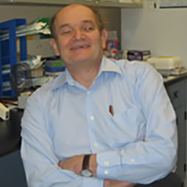
David Hedley
MD, Leeds (UK)

At A Glance
- Focus on cancer biology and treatment; emphasis on pancreatic cancer which is highly aggressive and difficult to treat.
- Lab makes extensive use of patient-derived xenografts that recapitulate the pathophysiology and genomic heterogeneity of pancreatic cancer patients.
- Internationally-recognized expertise in development and application of advanced flow cytometry techniques for studying cancer biology.
- Investigates novel, mechanism-based approaches to individualized treatment.
- Extensive network of collaborations involving clinical programs, pathology, and basic research.
Short Bio
Dr Hedley trained in internal medicine and medical oncology in the UK, including a three year thesis project on tumour immunology at the Royal Marsden Hospital/Institute for Cancer Research, University of London. From 1981-89 he was junior faculty at the University of Sydney, Australia, with staff positions as medical oncologist, Royal Prince Alfred Hospital, Sydney, and head of the flow cytometry laboratory, Ludwig Institute for Cancer Research. In 1990 he was appointed as a clinician/scientist in medical oncology at Princess Margaret Hospital, and senior scientist, Ontario Cancer Institute, with the rank of associate professor of medicine, University of Toronto. Since 1998 he has been a full professor, with cross appointments in the Departments of Medical Biophysics and Laboratory Medicine and Pathobiology. His major laboratory focus is on cancers arising in the pancreas and biliary tract, and aligns closely to the clinic in order to understand the high lethality of these cancers, and to explore novel approaches to treatment.
Research Synopsis
Preclinical and early clinical development of molecular cancer therapeutics. My main goal is the development of new, more effective forms of cancer treatment. This effort involves bridging the divide between basic science, pathology, and clinical oncology, and I consider my wide breadth of knowledge and experience at the interface between clinical medicine and basic science, and my ability to form effective collaborations across a broad range of disciplines, to be my strongest personal attributes. For the past 15 years my main focus has been on pancreatic cancer. My laboratory was one of the first to make use of primary xenografts to study cancer biology and molecular therapeutics. Utilizing my expertise in cellular analysis based on flow cytometry and advanced fluorescence imaging, I have been able to develop sophisticated approaches for studying complex biology in human cancers, and then use this to examine how novel agents are working at the molecular level. I have a particular interest understanding the mechanisms of drug resistance in pancreatic cancers, and in the way responses to tumour microenvironments alter the response to anticancer agents.
Understanding the microenvironment of pancreatic cancer. Hypoxia develops during solid tumour growth, and high levels are associated with metastasis development and resistance to standard treatments. We noted a wide range in the levels of hypoxia in our primary xenografts, and a powerful association with aggressive growth and metastasis. Our ongoing studies measuring hypoxia directly in patients confirms the wide heterogeneity in the levels of hypoxia, and by late 2016 we expect to confirm if this is associated with poor patient outcome. The patient tumours are also undergoing whole genome sequencing, as well as sophisticated tissue analysis using imaging mass cytometry, allowing us to probe deeply into the underlying mechanisms of hypoxia development in pancreatic cancer, and to search for potential drug targets for testing in patients. Our laboratory has established over 40 primary xenografts from this patient cohort, allowing us to explore mechanism-based treatment in the lab linked directly to the patient.
Development of Analytical Platforms based on Flow Cytometry and Imaging for Clinical Oncology. I have more than 30 years of experience in the field of flow cytometry, and am internationally recognized for the development of many novel techniques for studying human cancer. I am currently investigating the development of high dimensional flow cytometry to study biological complexity in pancreatic cancer, with particular emphasis on tumour hypoxia, cell signalling, damage response, and genomic instability. My laboratory also develops slide-based techniques for application to histological sections. Important features include the use of multiple fluorescence probes to allow co-localization of several molecules. Most recently we have been collaborating with Fluidigm Canada on the development of the revolutionary platform Imaging Mass Cytometry (IMC). Using antibody probes labelled with stable isotopes from the lanthanide series and pulsed UV laser ablation couple to time of flight mass spectrometry, IMC is capable of imaging 40+ antibodies on histological specimens at a resolution similar to x20 objective light microscopy. We are developing this technique for high content phenotypic analysis of pancreatic cancers, linked to our longstanding interest in the biology of hypoxia, and in experimental therapeutics.
Recent Publications
- Chang Q, Chandrashekhar M, Ketela T, Fedyshyn Y, Moffat J, Hedley D. Cytokinetic effects of Wee1 disruption in pancreatic cancer. Cell Cycle 2016; 15(4):593-604.
- Chang Q, Hedley D. Emerging applications of flow cytometry in solid tumor biology. Methods 2012; 57(3):359-67.
- Chang Q, Jurisica I, Do T, Hedley DW. Hypoxia predicts aggressive growth and spontaneous metastasis formation from orthotopically-grown primary xenografts of human pancreatic cancer. Cancer Res 2011.
- Dhani NC, Serra S, Pintilie M, Schwock J, Xu J, Gallinger S et al. Analysis of the intra- and intertumoral heterogeneity of hypoxia in pancreatic cancer patients receiving the nitroimidazole tracer pimonidazole. Br J Cancer 2015; 113(6):864-71.
- Lohse I, Borgida A, Cao P, Cheung M, Pintilie M, Bianco T et al. BRCA1 and BRCA2 mutations sensitize to chemotherapy in patient-derived pancreatic cancer xenografts. Br J Cancer 2015; 113(3):425-32.
- Mamaghani S, Simpson CD, Cao PM, Cheung M, Chow S, Bandarchi B et al. Glycogen Synthase Kinase-3 Inhibition Sensitizes Pancreatic Cancer Cells to TRAIL-Induced Apoptosis. PLoS One 2012; 7(7):e41102.
- Metran-Nascente C, Yeung I, Vines DC, Metser U, Dhani NC, Green D et al. Measurement of Tumor Hypoxia in Patients with Advanced Pancreatic Cancer Based on 18F-Fluoroazomyin Arabinoside Uptake. J Nucl Med 2016; 57(3):361-6.
