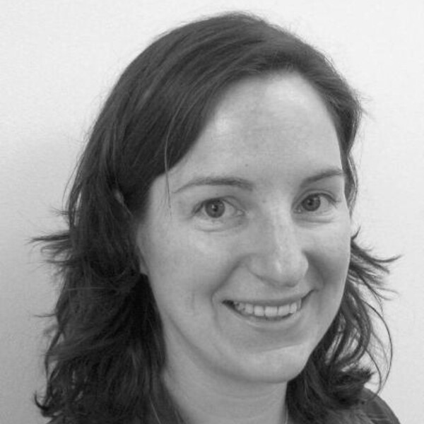
Christine Démoré
PhD, Queen's University

Short Bio
Christine Démoré completed her BSc Engineering Physics in 2000 and PhD Physics in 2006, both at Queen’s University, Kingston, Canada, before moving to Scotland. From 2007 to 2015 she was based at the Institute for Medical Science and Technology, University of Dundee, and at the School of Engineering, University of Glasgow in 2016. Dr. Démoré is now a Scientist at Sunnybrook Research Institute, and Assistant Professor in the Department of Medical Biophysics, University of Toronto. From 2013 to 2016 she held the Royal Society of Edinburgh / Caledonian Research Fund Biomedical Personal Research Fellowship, and she was awarded the IEEE Ultrasonics Young Investigator Award in 2015.
Research Synopsis
My research involves the development and application of ultrasonic devices and systems tailored for emerging biomedical applications. My work includes developing new ultrasound probes for a range of medical imaging applications, and creating devices for acoustic tweezing with applications in biosciences research.
The primary focus of my research is the development of miniature, high resolution ultrasound for clinical imaging. The ultrasound scanners commonly used in most clinical situations can provide images with resolution on the order of 1 mm. While this is useful for many clinical applications, there are many clinical and pre-clinical applications that can benefit from improved image resolution. Micro-ultrasound, with resolution in the range of 10 – 100 μm, is needed to resolve the fine structure of tissues, requiring in turn new ultrasound probes, or transducer arrays, capable of imaging with these resolutions to enable the new clinical applications.
Since both the probe dimensions and image resolution scale with the imaging wavelength (and conversely with operating frequency), miniaturised probes can to be developed. This introduces many challenges in fabricating new devices, but also creates opportunities for integrating miniature probes in endoscopes or other interventional tools like biopsy needles, that can be used for diagnosis and guiding a range of clinical procedures. Examples are the design of new probes integrated into biopsy needles optimised for guiding procedures in neurosurgery, or integrated into a swallowable pill for investigating gastrointestinal diseases. Alongside the development of new devices, my group is exploring the clinical and pre-clinical applications of micro-ultrasound imaging.
I have also developed transducer arrays for biosciences research applications, creating arrays with independently controlled elements that generate reconfigurable ultrasound fields for trapping and moving particles and cells. I have also used reconfigurable arrays to produce complex ultrasound fields, demonstrating real-world versions of science fiction concepts such as “Sonic Screwdrivers” and “Tractor Beams”.
Recent Publications
1. J. Yang, E. Chérin, J. Yin, I.G. Newsome, T. Kieski, G. Pang, C. Carnevale, P. A. Dayton, F.S. Foster, C.E.M. Démoré, “Characterization of an Array-Based Dual Frequency Transducer for Superharmonic Contrast Imaging,” IEEE Transactions on Ultrasonics, Ferroelectrics, and Frequency Control, Vol 68(7), p. 2419-2431, July 2021. (Trainee publication: Principal supervisor) [Senior Responsible Author]
Presents the acoustic performance of a dual-frequency transducer array with a 2 MHz array integrated behind a 20 MHz array; demonstrates superharmonic contrast imaging performance both in vitro and in vivo; represents an important step towards real-time superharmonic contrast imaging with interleaved B-mode imaging.
2. A. Chandra, Y. Soenjaya, J. Yan, P. Felts, G. McLeod, C.E.M. Démoré, “Real-time visualisation of peripheral nerve trauma during subepineural injection in pig brachial plexus using micro-ultrasound,” British Journal of Anaesthsia, Vol 127 (1), p.153-163, Jul 2021. (Trainee publication: Principal supervisor) [Co- Senior Responsible Author]
Reports real-time imaging peripheral nerve anaesthetic blocks, with comparison to histology, to understand morphological changes in nerves during extraneural and intraneural injections; gives insight into the physical response of nerves to external forces; shows that nerves translate and rotate, and fascicles within nerve translate and rotated in response of nerves to needle insertion and intraneural injection.
3. G. McLeod, S. Zhang, A. Sadler, A. Chandra, P. Qiao, Z. Huang, C.E.M. Démoré, “Validation of the Soft Embalmed Thiel Cadaver as a High Fidelity Simulator of Pressure during Targeted Nerve Injection,” Regional Anaesthesia and Pain Medicine , Vol 46(6), June 2021. [Co-Senior Responsible Author]
Presents measurement of extraneural and intraneural injection pressure during ultrasound-guided peripheral anaesthetic blocks on cadaveric models, demonstrating similar injection pressures to literature and in vivo porcine models, and supporting Thiel-embalmed cadavers as suitable simulators for training and research in peripheral anaesthesia.
4. J. Kenny, C.E. Munding, J.K. Eibl, A.M. Eibl, B.F. Long, A. Boyes, J. Yin, P. Verecchia, M. Parotta, R. Gatzke, P.A. Magnin, P.N. Burns, F.S. Foster, C.E.M. Démoré, “A Novel Hands-free Ultrasound Patch for Monitoring Quantitative Doppler in the Carotid Artery,” Scientific Reports, Vol 11(1), p. 7780, Apr 8 2021. [Senior Responsible Author]
Reports the design and early clinical demonstration of the FloPatch, the first wireless, wearable continuous-wave Doppler ultrasound patch that adheres to the neck and tracks Doppler blood flow metrics in the common carotid artery using an automated algorithm.
5. E. Cherin, J. Yin, A. Forbrich, C. White, P.A Dayton, F. S Foster, C.E.M. Démoré, “In vitro superharmonic contrast imaging using a hybrid dual frequency probe,” Ultrasound in Medicine and Biology, vol. 45(9), p. 2525-2539, May 2019. [Senior Responsible Author]
Reports on and investigates superharmonic contrast imaging using two 1.7 MHz transducers positioned along each side of a 20 MHz commercial micro-ultrasound array, representing an informative step toward array-based dual-frequency imaging probes designed for specific applications.
Graduate Students
Felipe Roa Garcia
Nidhi Singh
Jing Yang
