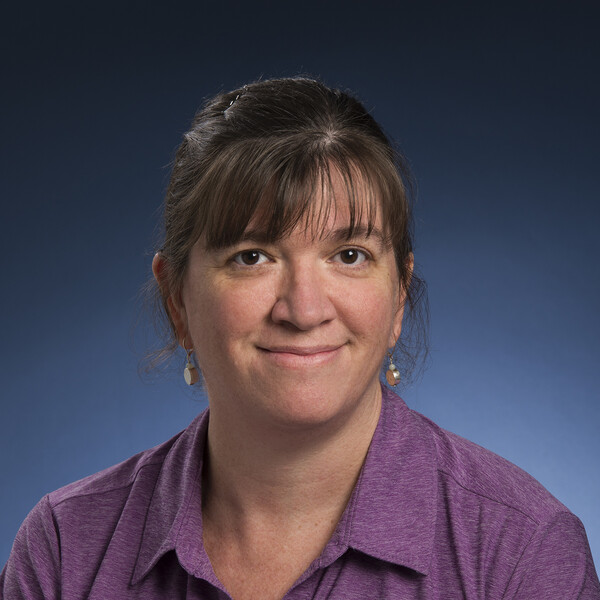
Laurie Ailles
PhD, University of British Columbia

Qualification
- Senior Research Scientist, Stanford University
- Research Associate, Stanford University
- Postdoctoral fellow, Pathology, Stanford University
- Postdoctoral fellow, Gene Therapy, Institute for Cancer Research, Candiolo, Italy
- PhD, Genetics, University of British Columbia
At A Glance
- Focus of the lab includes cancer biomarker discovery, cancer-associated fibroblasts, cancer mechanisms and epigenetics
- All work is done using primary patient-derived cancer tissue specimens, as well as patient-derived primary cultures and xenografts
- Diseases studied include head and neck squamous cell carcinoma, high-grade serous ovarian cancer and clear cell renal cell carcinoma
- We have established a “living biobank” of patient-derived xenografts that can be used to assay cancer stem cells, evaluate drug responses and development of drug resistance, and to perform a wide range of novel, clinically relevant studies.
- We collaborate extensively with other labs, clinicians and clinician scientists.
Research Synopsis
Identification of prognostic biomarkers and novel targets for head and neck squamous cell carcinoma (HNSCC) patients
Individualized treatment decisions are difficult in HNSCC due to the lack of prognostic and predictive biomarkers. Poor HNSCC survival rates clearly indicate a need for new and better therapies. Dr. Ailles and her team are working to identify prognostic biomarkers for patient stratification in HNSCC using genomic characterization of primary tumor tissues in combination with their functional behavior in tumor engraftment assays in immune-compromised mice. They are focused on identifying novel targets, pathways, and candidate drugs for the treatment of the most aggressive HNSCC tumors using patient-derived xenografts.
Epigenetics in clear cell renal cell carcinoma (ccRCC)
The Ailles team uses primary patient-derived cultures and xenografts to study the epigenetic deregulation occurring in ccRCC. More specifically, they focus on identifying the downstream effects on target genes and pathways and identifying small molecules with rapid clinical translational potential.
Targeting cancer-associated fibroblasts (CAFs) in solid tumors
Cancer-associated fibroblasts (CAFs) are known to play a role in promoting cancer proliferation, invasion, and chemo-resistance through acquisition of an “activated” state. The Ailles team has developed methods for the isolation of CAFs from primary patient-derived tumors and carried out molecular characterization of these tumours to identify molecular interactions between CAFs and cancer cells that may represent therapeutic targets. They have demonstrated functional heterogeneity within the CAF population, and are continuing to investigate strategies for epigenetic reprogramming of CAFs to a less activated state.
For more information, please see the ailleslab website.
Recent Publications
- Hussain A, Voisin V, Poon S . . . Tang KH, Paterson J, Clarke B, Bernardini MQ, Bader G, Neel B, Ailles L. Distinct fibroblast functional states drive clinical outcomes in ovarian cancer and are regulated by TCF21. Journal of Experimental Medicine, 217: e20191094, 2020.
- Ruicci K, Meens J, Sun RX . . . Mymryk JS, Barrett JW, Boutros P, Ailles L*, Nichols AC*. A controlled trial of HNSCC patient-derived xenografts reveals broad efficacy of PIK3CA inhibition in controlling tumor growth. International Journal of Cancer, 145: 2100-2106, 2019.
- Sinha A, Hussain A, Ignatchenko V . . . Ho VWH, Neel BG, Clarke B, Bernardini MQ, Ailles L, Kislinger T. N-Glycoproteomics of patient-derived xenografts: A strategy to discovery tumor-associated proteins in high-grade serous ovarian cancer. Cell Systems, 8: 345-351, 2019.
- Karamboulas C, Bruce J, Hope AJ . . . Tiedemann R, Liu F-F, Bratman SV, Pugh T, Xu W, Ailles L. Patient-derived xenografts for prognostication and interrogation of precision medicine in head and neck squamous cell carcinoma patients. Cell Reports, 25:1318-1331, 2018.
- Lobo N, Gedye C, Apostoli A, Stickle N, Hyatt E, Evans A, Jewett MAS, Ailles L. Efficient generation of patient-matched malignant and normal primary cell cultures from clear cell renal cell carcinoma patients: clinically relevant models for research and personalized medicine. BMC Cancer, 16:485-499, 2016.
- Gedye C, Sirskyj D, Lobo N . . . Kulkarni G, Zlotta A, Evans A, Finelli A, Jewett MAS, Ailles LE. Standard experimental methods substantially underestimate renal carcinoma initiating cell frequencies. Scientific Reports, 6:25220, 2016.
Graduate Students
Baljit Kaur
Stephanie Poon
Joe Walton
